
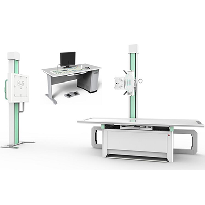
High Frequency Digital X-Ray Radiography System AM-AR-7300D
Main Features
AR-7300D high frequency digital x-ray equipment can meet different parts radiography, such as head, chest, abdomen, waist, lumber, thoracic, pelvis, limbs etc. And meet different positions radiography, such as lay decubitus, normotopia, side position etc. it is fixed digital x-ray machine and we can use it at bus and truck.
9 people are viewing this product right now

- Main Technical Parameters:
| Item | Content | Technical Parameters |
| Power | Voltage | 380V±10% |
| Frequency | 50Hz±1Hz | |
| Capacity | ≥65kVA | |
| Internal Resistance | ≤0.17Ω | |
| High Frequency High Voltage X-Ray Generator | Power | 56KW |
| Inverter Frequency | 440 KHz | |
| Tube voltage | 40kV—150kV ,continuous adjustment | |
| Tube current | 10mA—710mA , grade adjustment | |
| Exposure time | 0.1ms—10000ms | |
| mAs | 0.1-900mAs | |
| X-ray source assembly
(TOSHIBA E7252X) |
Tube Focus:big/small | 1.2mm /0.6mm |
| Input power | Big focus 75kW Small focus 27kW | |
| Anode thermal capacity | 210KJ | |
| Rotary anode speed | 9700rpm | |
| Digital Flat Panel Detector | Active area | 427(H)×427(V) |
| Pixel matrix: | 3072(H)×3072(V) | |
| Pixel pitch: | 139 μm | |
| Cycle time: | Less than 6 s | |
| Limiting resolution | 3.1 lp/mm typ | |
| A / D transition: | 14 bit | |
| Energy range | 40 – 150 kVp | |
| Maximum entrance dose (low gain) | 4 mR / frame | |
| power Input: | DC 24V2A | |
| Bucky Stand | Up and down movement | Not less than 1300mm |
| Table | SID | 500~1280mm |
| Table bearing | 135kg |
- Original imported high frequency host and high speed tube
-TOSHIBA highest inverter frequency x-ray tube and high frequency voltage generator.
Constant direct current high voltage output, it can achieve high quality monochromatic X-Ray and eliminate the harmful effect of soft ray to image thoroughly.
-Equipped with high performance and large capacity X-Ray tube
Adopt 0.6/1.2mm2 dual focus, 300KHU large capacity and high speed X-Ray tube,it is suitable for long time and high intensity clinical examination.
-Intelligent high voltage control system with multiple human APR, LCD display show APR rules, and set according patients figure, position and part automatically, it’s fast and convenient.
-Computer progress control, remote operate, electric multi-lead collimator, adjust X-ray range as diagnosis need, this reduce X-ray harm for patients and atmosphere
-Multi self-diagnosis program, has RS232 port, and with error indication function.
- High definition digital image chain to ensure that minor lesions can not be passed.
-Imported DR flat panel detector, it does not need any conversion but it can finish digital photography of each position to achieve real direct digital.
-DR flat panel detector DR connected with image processor achieves low noise and rich contrast image, edge enhancement filtering device makes the edge of image more clear and sharp.
-3k×3k gathering matrix, more than 900 mega pixel, promise high photo space definition, and good signal-no-noise ratio.
-17”*17” fixed flat panel detector can cover whole abdomen and whole chest, no limitation for photography.
-Fast image speed, work together with DROC soft, and promote work efficiency.
-A-si stable function, low requirement for temperature, and no crystal under high temperature.
- Equipped with new digital image system achieves rich digital image processing function
Advanced digital image processing function
-Pre-assembled Windows professional operating system and professional image processing software
Image playback: thumbnails view, sequence replay tools.
Image processing: W/L adjustment, arrows and words etc labels, angle and distance measurement, image scaling, translation, up/down conversion, left/right conversion, rotation, black and white reversal.
-Image storage: image real-time storage, DICOM image send, A shampoo Burning Studio, derived storage (choose various storage ways Bitmap, JPEG, AVI etc to be used in Word and Power-point office software, it is convenient for the doctors to write diagnostic reports and papers ).
-DICOM3.0: can be connected with laser camera to print film and PACS NETWORK.
-Medical record management: database management, graphic report, support for WORKLIST.
- DR Workstation
Touch screen operation and UPS for the workstation
Computer system:
B150 PLUS motherboard; processor: Intel Core I5 6400
Memory: 8 GB DDR3
Hard disk: 1T SATA (7200 rpm)
Optical drive: DVR-221CHV CD burner
NIC: Intel(R)gigabit CT
Graphics card: gtx1050
Display: Dell (DELL) U2412Mc display


Reviews
There are no reviews yet.